Research
 |
 |
 |
1. Mapping longitudinal development and hemispheric asymmetries of cortical thickness in infants
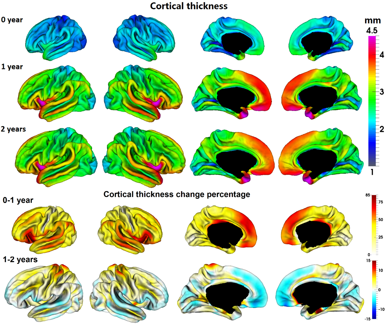 |
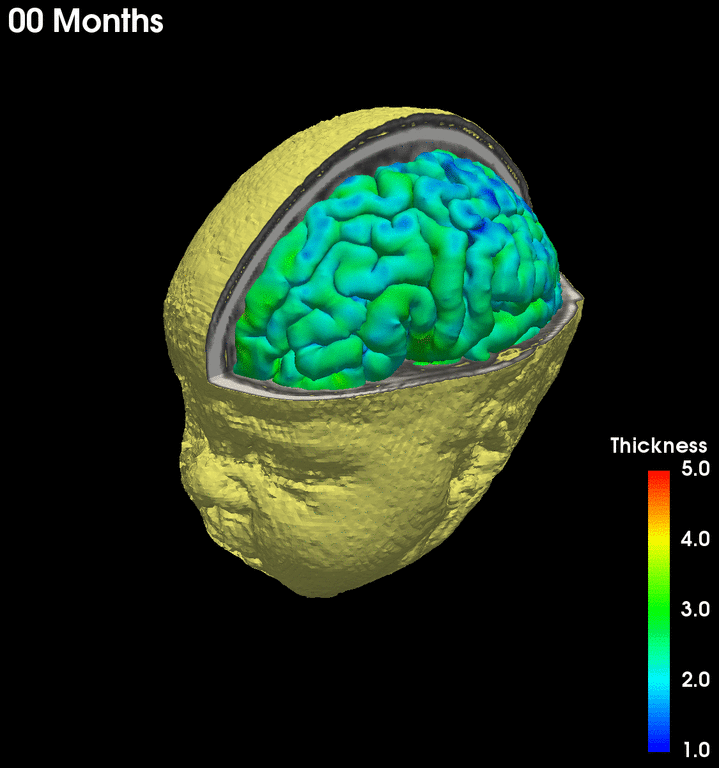
Cortical thickness (CT) is related to normal development and neurodevelopmental disorders. It remains largely unclear how the characteristic patterns of CT evolve in the first 2 years. We systematically characterized for the first time the detailed vertex-wise patterns of spatial distribution, longitudinal development, and hemispheric asymmetries of CT at 0, 1, and 2 years of age, via surface-based analysis of 219 longitudinal MRI scans from 73 infants. Spatial patterns, longitudinal development, and hemispheric asymmetries of cortical thickness in infants from birth to 2 years of age. Gang Li, Weili Lin, John H. Gilmore, Dinggang Shen. The Journal of Neuroscience, vol. 35, pp. 9150-9162, 2015. |
2. Learning-based predicting infant cortical surface development
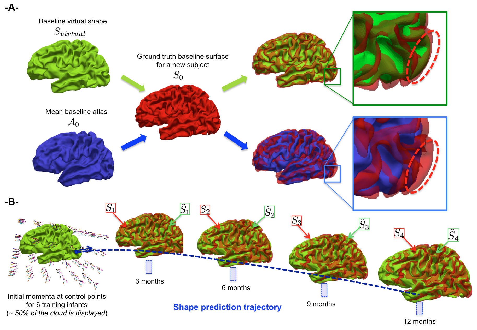 |
Longitudinal neuroimaging analysis methods have remarkably advanced our understanding of early postnatal brain development. However, learning predictive models to trace forth the evolution trajectories of both normal and abnormal cortical shapes remains broadly absent. To fill this critical gap, we pioneered the first prediction model for longitudinal developing cortical surfaces in infants using a spatiotemporal varifold-based learning framework solely from the baseline cortical surface. Predicting infant cortical surface development using a 4D varifold-based learning framework and local topography-based shape morphing. Islem Rekik, Gang Li, Weili Lin, Dinggang Shen. Medical Image Analysis, vol. 28, pp. 1-12, 2016. |
3. Mapping cortical structural connectivity development in infants
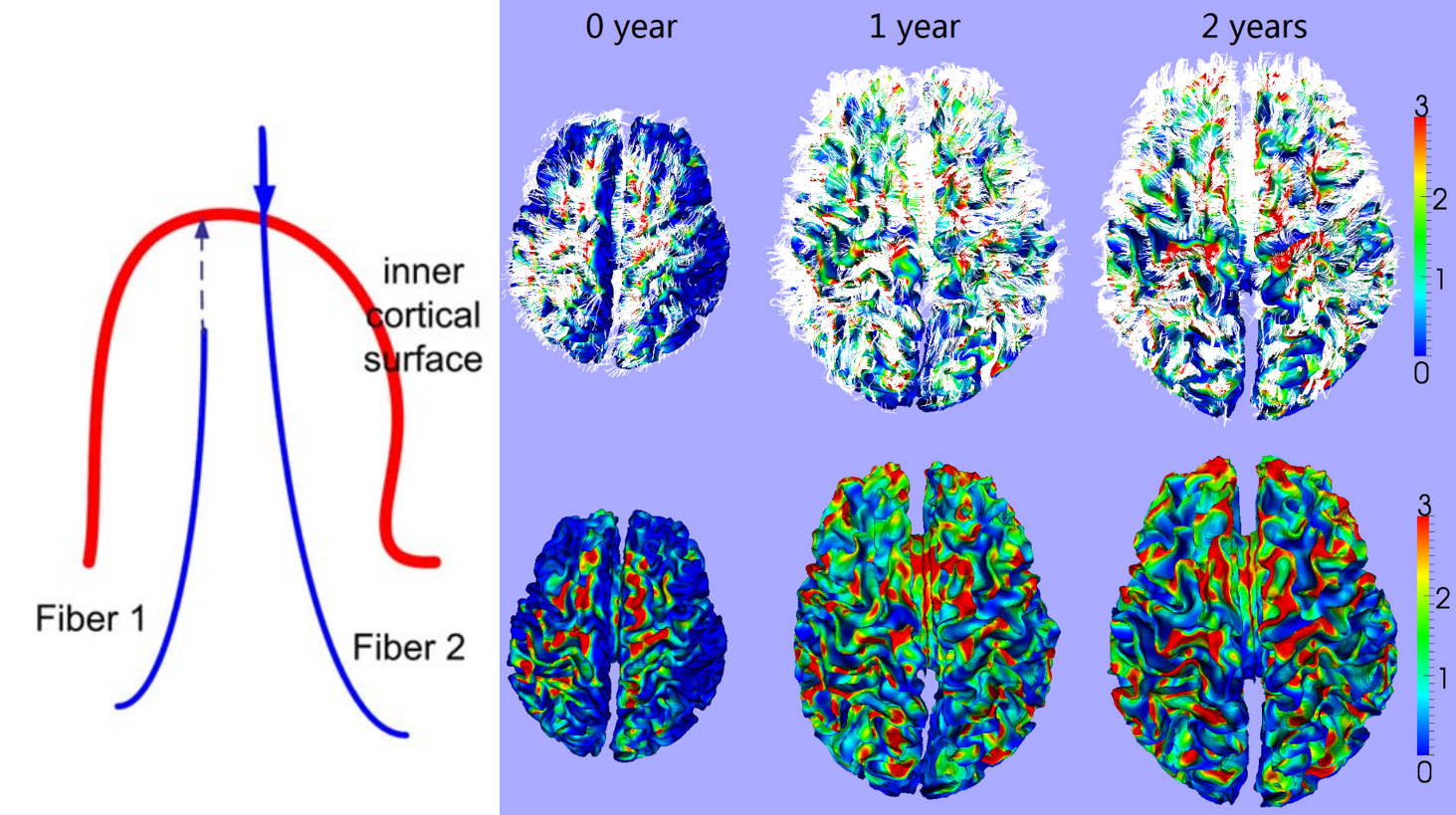 |
Quantitatively characterizing the development of cortical anatomical networks during the early stage of life plays an important role in revealing the relationship between cortical structural connection and high-level functional development. The development of correlation networks of cortical-thickness, cortical folding, and fiber-density is systematically analyzed in this article to study the relationship between different anatomical properties during the first 2 years of life. Longitudinal development of cortical thickness, folding, and fiber density networks in the first 2 years of life. Jingxin Nie, Gang Li, Li Wang, Feng Shi, Weili Lin, John H. Gilmore, Dinggang Shen. Human Brain Mapping, vol. 35, pp. 3726–3737, 2014. Spatiotemporal patterns of cortical fiber density in developing infants, and their relationship with cortical thickness. Gang Li, Tianming Liu, Dong Ni, Weili Lin, John H. Gilmore, Dinggang Shen. Human Brain Mapping, vol. 36, pp. 5183-5195, 2015. |
4. Mapping spatio-temporal patterns of deep sulcal landmarks on infant cortical surfaces
 |
Sulcal pits, the locally deepest points in sulci of the highly convoluted cerebral cortex, are found to be spatially consistent across human adult individuals. Little is known about the spatial distribution and temporal development of sulcal pits in the first 2 years of life, which is the most dynamic and critical period of postnatal brain development. We systemically investigated the spatial distribution and temporal development of sulcal pits in major cortical sulci from 73 healthy infants. Our results suggest that the spatially consistent distributions of sulcal pits in major sulci across individuals have already existed at term birth and this spatial distribution pattern keeps relatively stable in the first 2 years of life, despite that the cerebral cortex expands dramatically and the sulcal depth increases considerably during this period. Spatial distribution and longitudinal development of deep cortical sulcal landmarks in infants. Yu Meng*, Gang Li*, Weili Lin, John H. Gilmore, Dinggang Shen. NeuroImage, vol. 100, pp. 206-218, 2014. (* Equal contribution) |
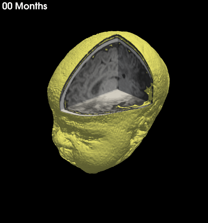
5. Consistent 4D reconstruction of longitudinal infant cortical surfaces
 |
This paper presents a method to reconstruct temporally-consistent cortical surfaces from longitudinal infant brain MR images, for accurate and consistent measurement of the dynamic cortex development in infants. Specifically, the longitudinal development of the inner cortical surface is first modeled by a deformable growth sheet with elasto-plasticity property to establish longitudinally smooth correspondences of the inner cortical surfaces. Then, the modeled longitudinal inner cortical surfaces are jointly deformed to locate both inner and outer cortical surfaces with a spatial–temporal deformable surface method. Measuring the dynamic longitudinal cortex development in infants by reconstruction of temporally consistent cortical surfaces. Gang Li, Jingxin Nie, Li Wang, Feng Shi, John H. Gilmore, Weili Lin, Dinggang Shen. NeuroImage, vol. 90, pp. 266-79, 2014. |
6. Mapping longitudinal development of cortical local gyrification (LGI) in infants
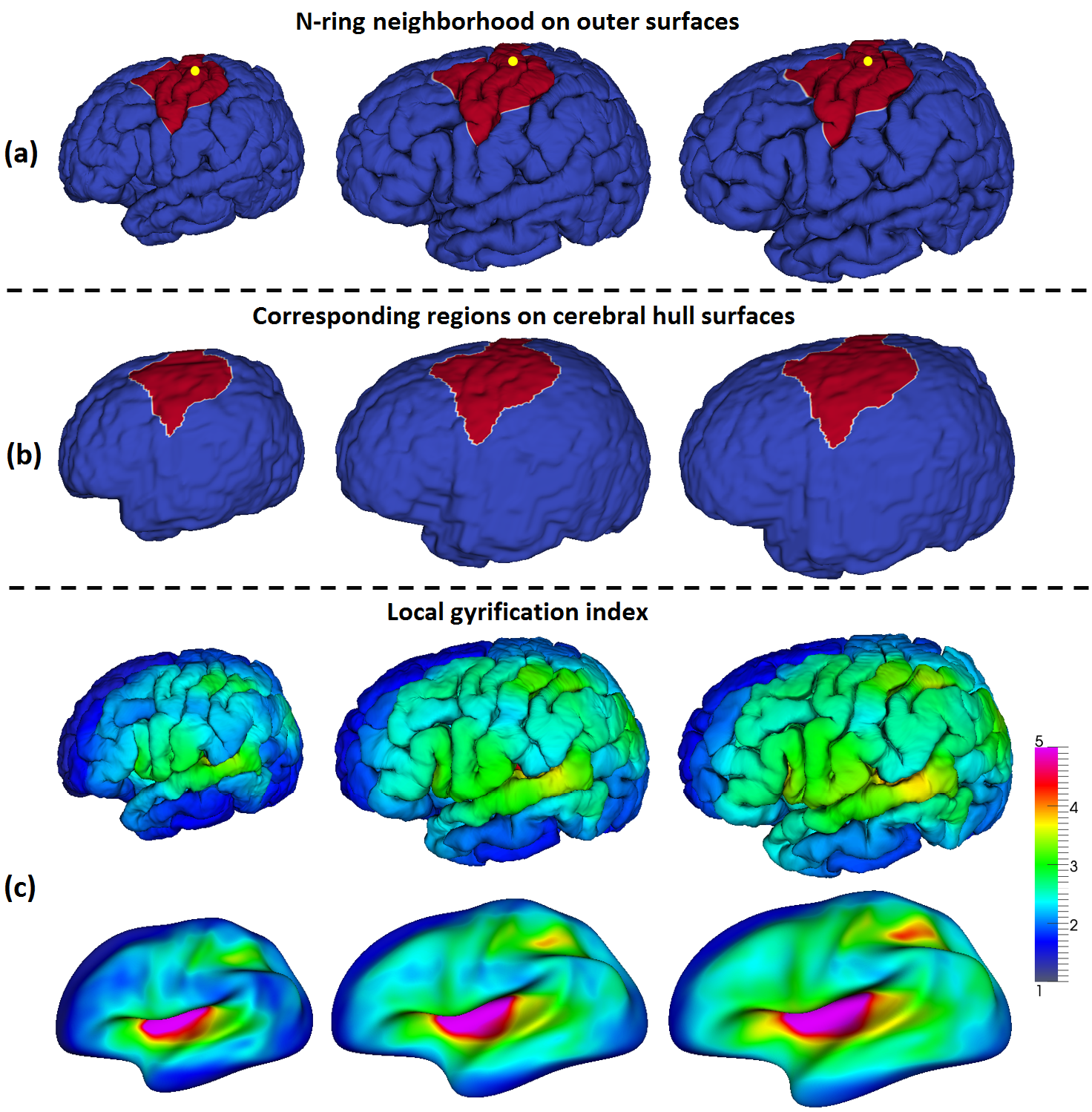 |
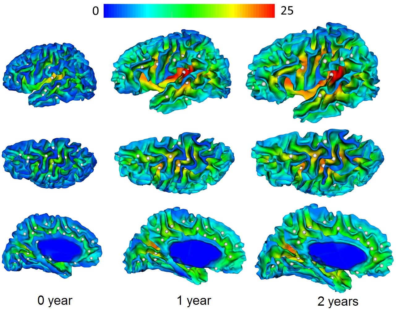
We developed a novel infant-specific method for mapping longitudinal development of local cortical gyrification in infants. By using this method, via 219 longitudinal 3T MRI scans from 73 healthy infants, we systemically characterized for the first time the longitudinal cortical global gyrification index (GI) and local GI (LGI) development in the first 2 years of life. We found that the cortical GI had age-related and marked development, with 16.1% increase in the first year and 6.6% increase in the second year. We also found marked and regionally heterogeneous LGI development in the first 2 years of life, with the high-growth regions located in the association cortex, whereas the low-growth regions located in sensorimotor, auditory, and visual cortices. Mapping longitudinal development of local cortical gyrification in infants from birth to 2 years of age. Gang Li, Li Wang, Feng Shi, Amanda E. Lyall, Weili Lin, John H. Gilmore, Dinggang Shen. The Journal of Neuroscience, vol. 34, pp. 4228-38, 2014. (Highlighted in the National Institute of Mental Health (NIMH)’s 2015-2020 Strategic Plan) |
7. Computational growth model for measuring dynamic cortical development in the first postnatal year
 |
Human cerebral cortex develops extremely fast in the first year of life. Quantitative measurement of cortical development during this early stage plays an important role in revealing the relationship between cortical structural and high-level functional development. This paper presents a computational growth model to simulate the dynamic development of the cerebral cortex from birth to 1 year old by modeling the cerebral cortex as a deformable elastoplasticity surface driven via a growth model. A computational growth model for measuring dynamic cortical development in the first year of life. Jingxin Nie, Gang Li, Li Wang, John H. Gilmore, Weili Lin, Dinggang Shen. Cerebral Cortex, vol. 22, pp. 2272-2284, 2012. A computational model of cerebral cortex folding. Jingxin Nie, Lei Guo, Gang Li, Tianming Liu. Journal of Theoretical Biology, vol. 264, pp. 467–478, 2010. (Reported by NIH/Stanford Biomedical Computation Review) |
8. Consistent and simultaneous labeling of longitudinal dynamic cortical surfaces in infants
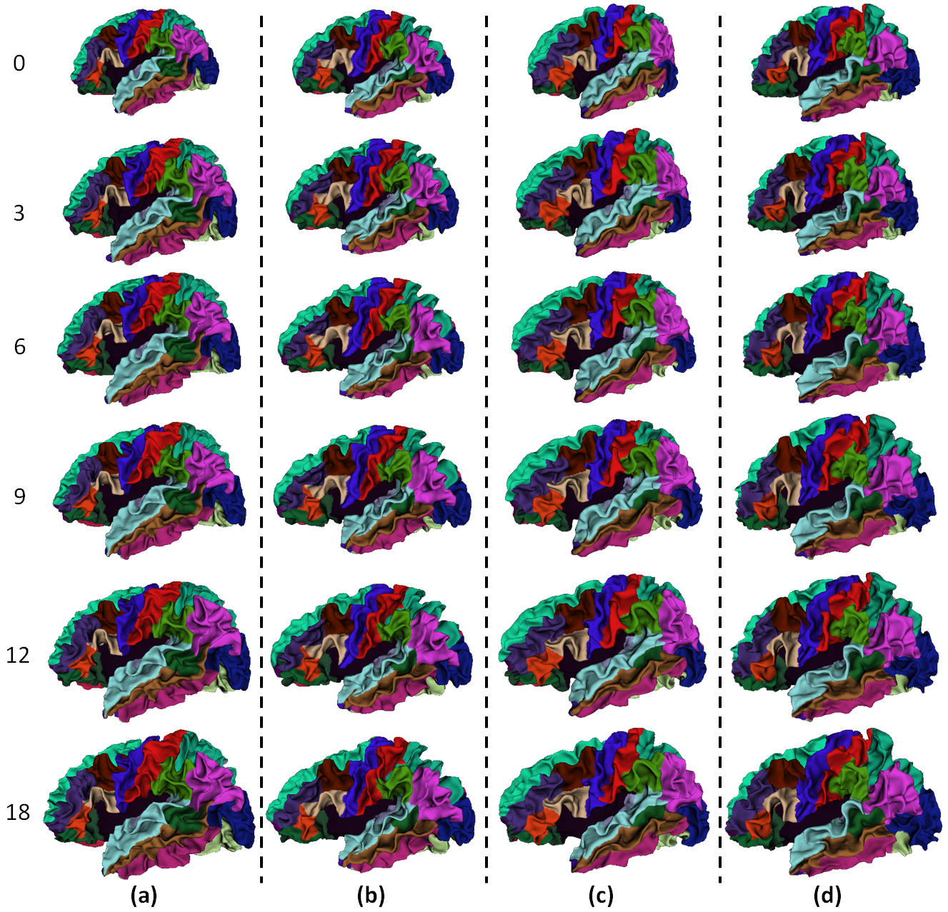 |
Accurate and consistent labeling of longitudinal cortical surfaces is essential to understand the early dynamic development of cortical structure and function in both normal and abnormal infant brains. We propose a novel method for simultaneous, consistent, and unbiased labeling of longitudinal dynamic cortical surfaces in the infant brain MR images. The proposed method is formulated as minimization of an energy function, which includes the data fitting, spatial smoothness and temporal consistency terms. Simultaneous and consistent labeling of longitudinal dynamic developing cortical surfaces in infants. Gang Li, Li Wang, Feng Shi, Weili Lin, Dinggang Shen. Medical Image Analysis, vol. 18, pp. 1274–1289, 2014. |
9. Mapping longitudinal hemispheric structural asymmetries of the cerebral cortex in infants
 |
Mapping cortical hemispheric asymmetries in infants increase our understanding of the origins and developmental trajectories of asymmetries. We analyze longitudinal cortical hemispheric asymmetries from birth to age 2 years using surface-based morphometry. Prominent asymmetries are found around the peri-Sylvian region and superior temporal sulcus at birth that evolve modestly from birth to age 2 years. Mapping longitudinal hemispheric structural asymmetries of the human cerebral cortex from birth to 2 years of age. Gang Li, Jingxin Nie, Li Wang, Feng Shi, Amanda E. Lyall, Weili Lin, John H. Gilmore, Dinggang Shen. Cerebral Cortex, vol. 24, pp. 1289-1300, 2014. |
10. Mapping longitudinal cortical surface area expansion in infants
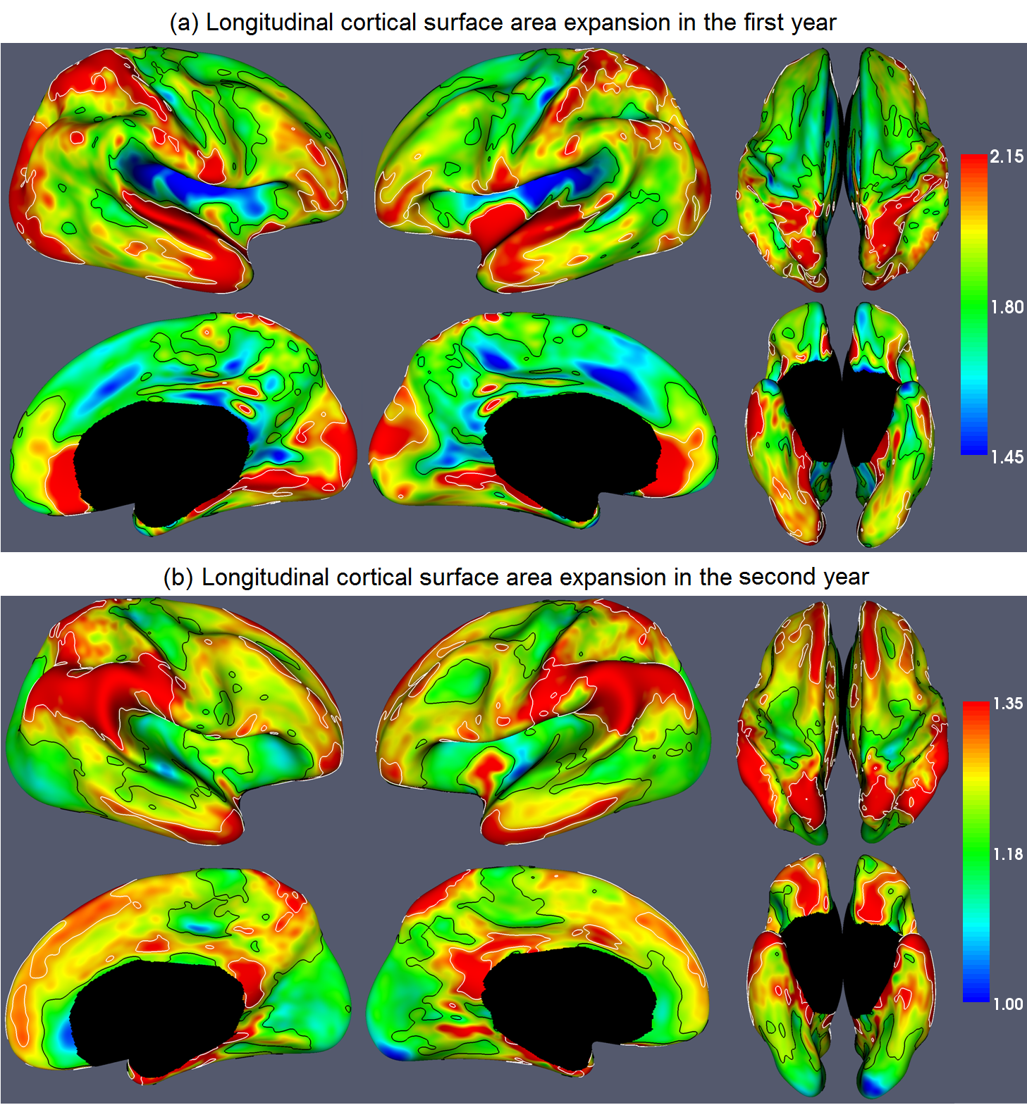 |
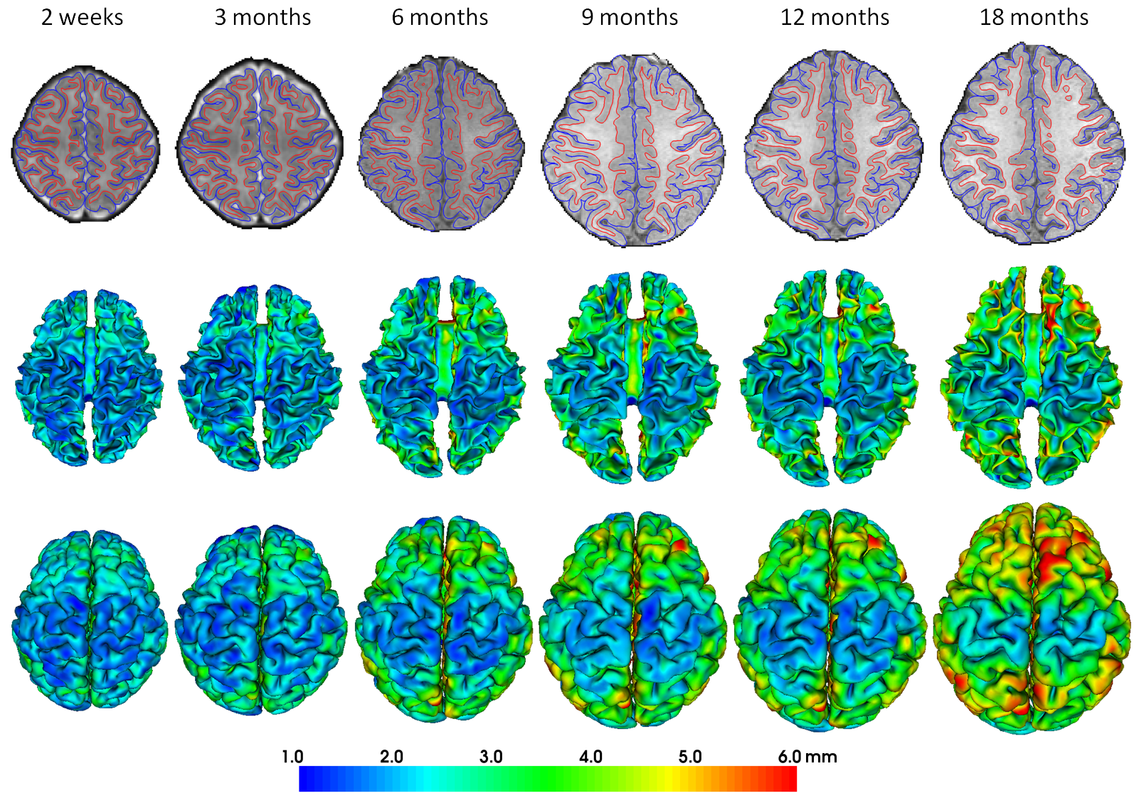
Human cerebral cortex develops dynamically in the first 2 years of life. Little is known about the longitudinal cortical surface expansion during this stage. We generate the first longitudinal surface-based atlases of human cortical structures at 0, 1, and 2 years of age from 73 healthy subjects, and study the longitudinal cortical surface expansion in the first 2 years of life. We find that cortical surface expansion is age related and region specific, which are related to cognitive and functional development at these stages. Mapping region-specific longitudinal cortical surface expansion from birth to 2 years of age. Gang Li, Jingxin Nie, Li Wang, Feng Shi, Weili Lin, John H. Gilmore, Dinggang Shen. Cerebral Cortex, vol. 23, pp. 2724-2733, 2013. |
11. Extraction of sulcal fundi curves on cortical surface
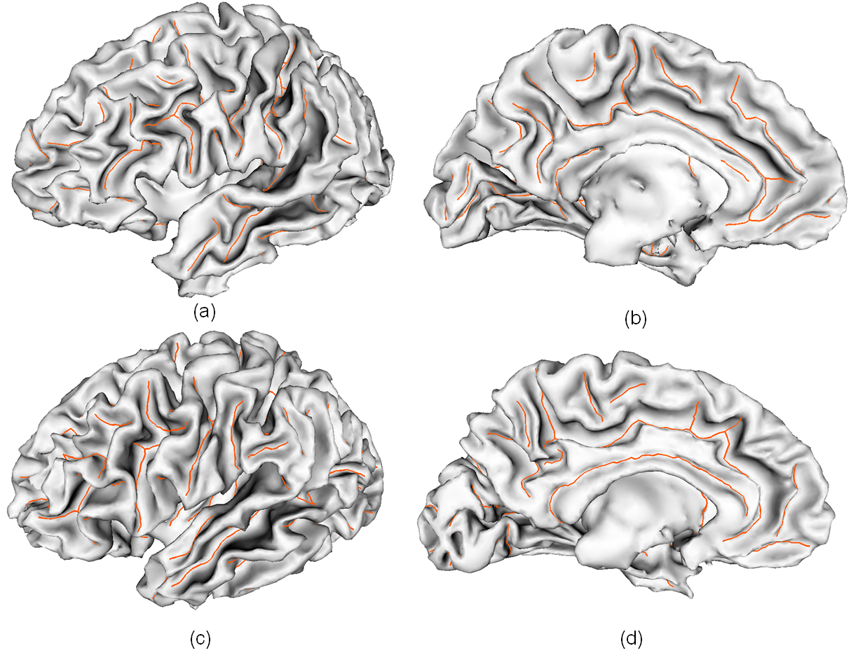 |
We propose a novel automated pipeline for extraction of sulcal fundi from triangulated cortical surfaces. The proposed method can find the accurate sulcal fundi using curvatures and curvature derivatives without any manual interaction. The extracted sucal fundi curves could be served as landmarks in brain image warping and used to analyze the variability of cortical folding. An automated pipeline for cortical sulcal fundi extraction. Gang Li, Lei Guo, Jingxin Nie, Tianming Liu. Medical Image Analysis, vol. 14, pp. 343-359, 2010. |
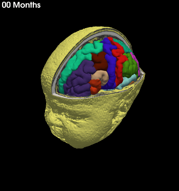
12. Consistent sulcal parcellation of longitudinal cortical surfaces
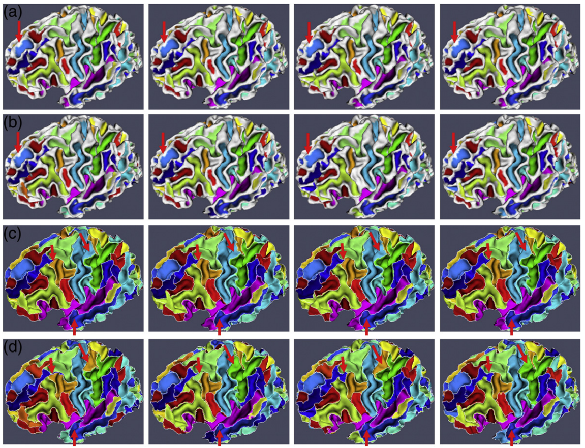 |
Accurate and consistent sulcal parcellation of longitudinal cortical surfaces is of great importance in studying longitudinal subtle morphological changes of adult human brains. Applying existing cross-sectional methods to longitudinal cortical surfaces might generate longitudinally-inconsistent results. We present a novel method for accurate and consistent sulcal parcellation of longitudinal cortical surfaces. Both spatial and temporal smoothness are imposed in the energy function to obtain consistent longitudinal sulcal parcellation results. Consistent sulcal parcellation of longitudinal cortical surfaces. Gang Li, Dinggang Shen. NeuroImage, vol. 57, pp. 76-88, 2011. |
13. Longituidnally consistent reconstruction of cortical surfaces from serial MRI
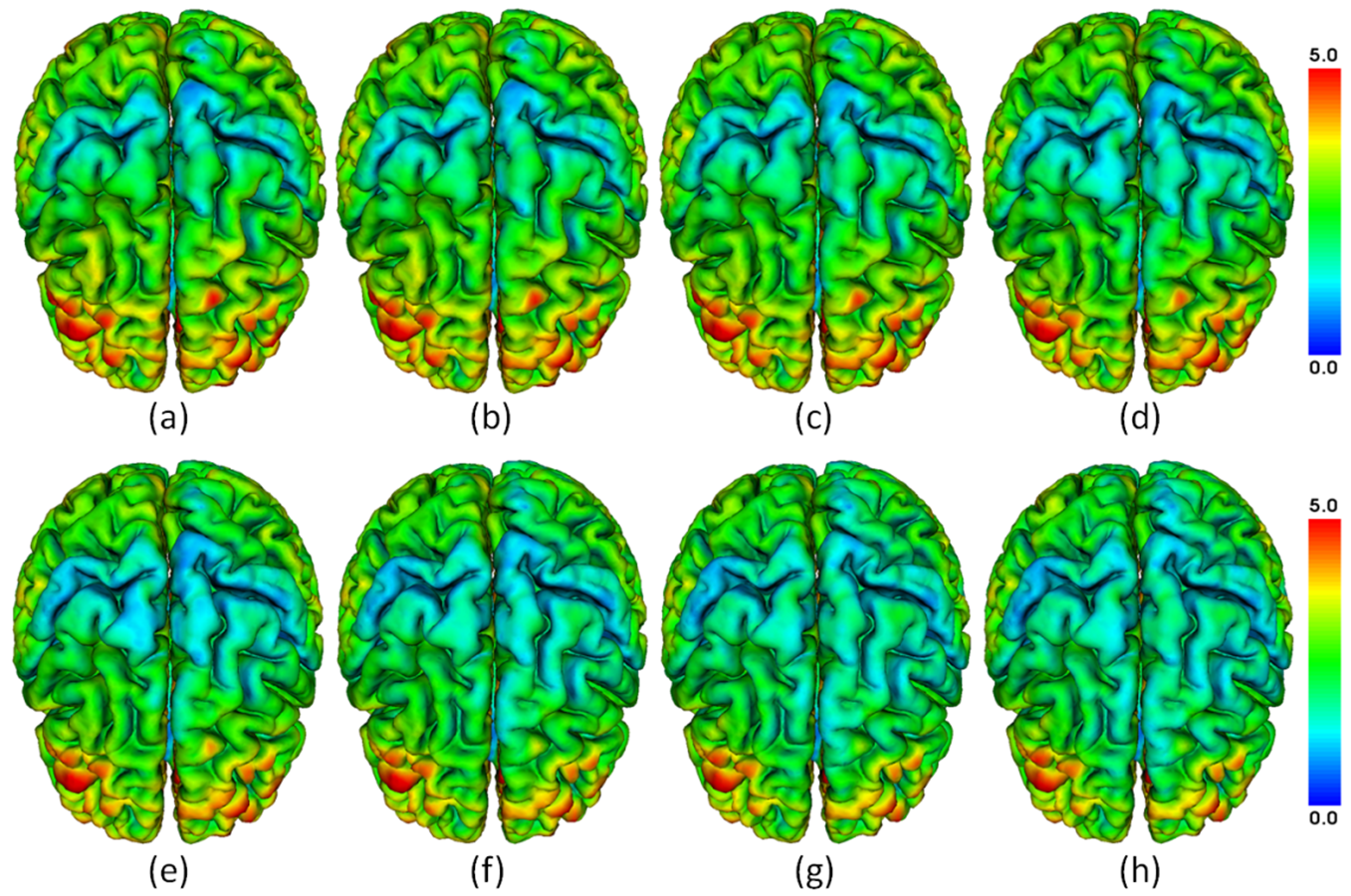 |
Consistent reconstruction of cortical surfaces from longitudinal human brain MR images is of great importance in studying longitudinal subtle change of the cerebral cortex. This paper presents a novel deformable surface method for consistent and accurate reconstruction of cortical surfaces from longitudinal brain MR images.The longitudinal cortical surfaces show overall decline trend of cortical thickness and its potential in distinguishing clinical groups. Consistent reconstruction of cortical surfaces from longitudinal brain MR images. Gang Li, Jingxin Nie, Guorong Wu, Yaping Wang, Dinggang Shen, ADNI. NeuroImage, vol. 59, pp. 3805-3820, 2012. |
14. Sulcal parcellation of individual cortical surface using flow tracking
 |
Automatic parcellation of the cortical surface into sulcal regions is of great importance in human rain mapping. A novel method is proposed for automatic cortical sulcal parcellation based on the geometric characteristics of cortical surface including its principal curvatures and principal directions. Automatic cortical sulcal parcellation based on surface principal direction flow field tracking. Gang Li, Lei Guo, Jingxin Nie, Tianming Liu. NeuroImage, vol. 46, pp. 923-937, 2009. |
15. Segmentation of 2D/3D densely touching cell nuclei in microscopy images
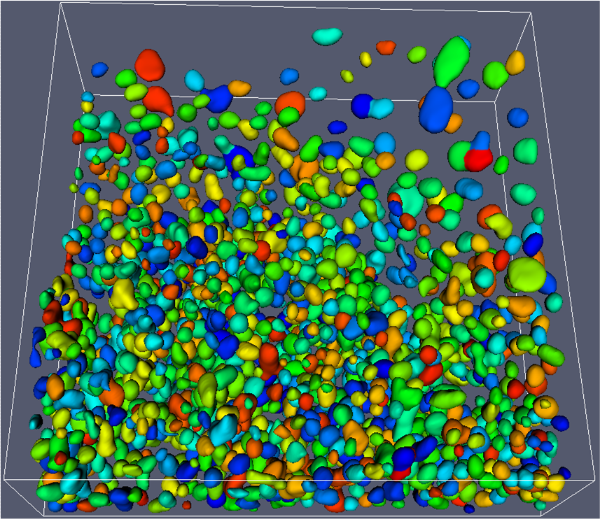 |
Reliable and accurate segmentation of cell nuclei from microscopic images is an important task in many computational biological studies. We present a novel, fully automated method for the segmentation of dense touching cell nuclei from 2D and 3D microscopic images, based on gradient flow field tracking and grouping. 3D cell nuclei segmentation based on gradient flow tracking. Gang Li, Tianming Liu, Ashley Tarokh, Jingxin Nie, Lei Guo, Andrew Mara, Scott Holley, Stephen TC Wong. BMC Cell Biology, 8:40, 2007. |
16. Bioimage informatics
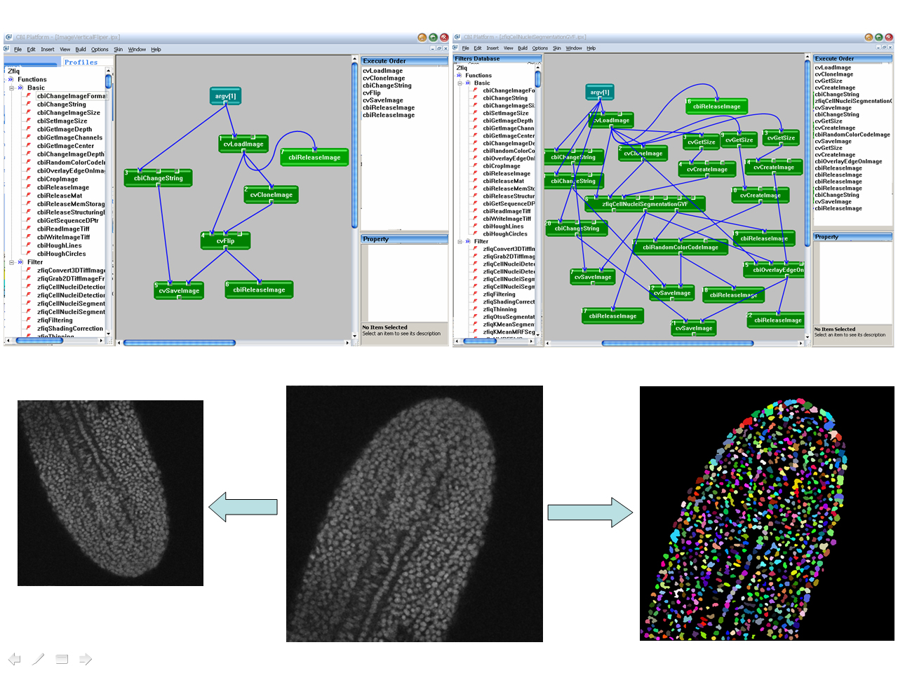 |
The tremendous amount of image data generated from microscopy has led to the bottleneck of data analysis and modeling. The zebrafish image quantitator (ZFIQ) software provides streamlined data processing and analysis capability for developmental biology and disease modeling. ZFIQ: a software package for zebrafish biology. Tianming Liu, Jingxin Nie, Gang Li, Lei Guo and Stephen TC Wong. Bioinformatics, vol. 24, pp. 438-439, 2008. (Software) Detection of blob objects in microscopy zebrafish images based on gradient vector diffusion. Gang Li, Tianming Liu, Jingxin Nie, Lei Guo, Jarema Malicki, Andrew Mara, Scott Holly, Weiming Xia, Stephen TC Wong. Cytometry Part A, vol. 71, pp. 835-845, 2007. (Cover Page) |